Project
protocol
—
Contents
Workflow
and
sampling
Equipment
Reagents,
supplies,
and
solutions
Definitions
Procedure
Data
References
Workflow
and
sampling
Workflow
Test |
Procedure
performed |
Equipment |
Age
(wk) |
Data
collected |
|
Mice
are
examined
visually
for
normal
health
and
appearance |
|
|
- |
2 |
Both
right
and
left
middle
ear
function
is
assessed |
tympanometer |
5-68 |
compliance,
volume,
gradient,
pressure |
3 |
Selected
mice
are
further
examined
for
ABR
(auditory
brainstem
response)
threshold
analysis |
|
|
ABR
threshold |
4 |
Selected
mice
with
abnormal
tympanogram
and
elevated
ABR
threshold
(PL/J)
are
confirmed
otoscopically |
video
otoscope |
5-68 |
video
images
of
ear
membranes |
5 |
Selected
mice
with
abnormal
tympanogram,
elevated
ABR
threshold
(LP/J),
and
otoscopic
images
are
necropsied
and
middle
ears
submitted
for
histology |
dissecting
kit |
|
- |
6 |
Histological
samples
are
stained
and
microscopically
examined
to
confirm
abnormal
tympanogram |
microscope |
5-68 |
|
Equipment
Tympanometer:
MT
10
(Interacoustics,
Assens,
Denmark)
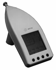
Figure
1.
A
hand-held
tympanometer.
Reagents,
supplies,
solutions
Anesthesia:
Avertin®
(Thribromoethanol)
5
mg/10g
BW
dose
Definitions
Acoustic
admittance:
The
degree
with
which
sound
waves
travel
through
the
eardrum
membrane.
Acoustic
compliance:
Synonymous
with
"acoustic
admittance".
Compliance:
degree
with
which
air
travels
(i.e.
determined
by
the
eardrum
and
the
middle
ear
system);
indicative
of
the
equivalent
volume
of
air
in
the
middle
ear.
Gradient:
refers
to
the
shape
or
width
of
the
tympanometer
curve.
Otosclerosis:
aberrant
bone
growth
of
the
middle
ear
resulting
in
structural
deficit
and
conductive
hearing
loss.
Pascal
(Pa):
unit
for
pressure
or
stress
where
1
Newton/m2
=
1
Pa.
Pressure:
refers
to
the
amount
of
air
pressure
applied
to
the
ear
canal
to
obtain
maximum
acoustic
or
eardrum
compliance.
Tympanogram:
resulting
chart
obtained
when
measuring
compliance
of
the
eardrum
using
a
tympanometer.
Three
general
types
of
tympanogram
tracings
have
been
described
in
the
literature.
A
normal
ear
gives
tracing
type
A
as
shown
in
Figure
2
below
(a
bell-shaped
curve
with
peak
admittance
occurring
at
or
near
0
daPa).
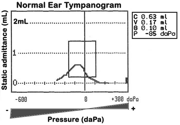
Figure
2.
An
example
of
a
normal
tympanogram
graphically
charting
compliance
(C
in
mL)
of
the
tympanic
membrane
under
changing
pressure
conditions.
The
equivalent
volume
(V
in
mL)
of
the
outer
ear
canal,
the
gradient
(G
in
mL)
and
the
pressure
in
dekapascal
units
(P
in
daPa)
at
maximum
compliance
are
also
given
on
the
right.
Tympanometry:
measurement
of
the
ability
of
the
eardrum
or
the
middle
ear
membrane
and
its
associated
bones
(hammer/malleus,
anvil/incus,
stirrup/stapes,
see
Figure
3
below)
to
transmit
sounds
in
the
form
of
pressure
waves.
When
subjected
to
changes
in
air
pressure,
the
intact
eardrum
stiffness
(impedance)
and
compliance
(admittance)
characteristics
can
be
thus
be
determined.
Volume:
refers
to
the
equivalent
volume
of
the
outer
ear
canal
with
reference
to
the
volume
in
the
middle
ear.
Acclimation
to
test
conditions
In
general
all
mice
are
acclimated
in
the
procedure
room
where
the
tympanometry
examinations
are
conducted.
Procedure
for
conducting
tympanometry
Pre-testing
preparations
a.
Tympanometry
is
conducted
in
a
quiet
animal
procedure
room.
Environmental
noise
is
maintained
at
a
minimum
of
50
decibels
sound
pressure
level
(dB
SPL).
b.
After
performing
a
comprehensive
calibration
of
the
sound
level
meter
and
bioacoustic
simulator,
a
mouse
is
then
prepared
and
given
short-term
anesthesia
intraperitoneally
(i.p.).
c.
Once
the
mouse
is
fully
anesthetized,
it
is
visually
and
quickly
examined
for
signs
of
developmental
defects
or
morphological
abnormalities.
The
external
ear
canal
is
checked
for
cerumen
(ear
wax)
or
debris
buildup
with
the
use
of
an
otoscope.
Any
potential
obstruction
to
the
ear
probe
opening
is
removed
including
excessive
ear
wax
and
hair.
d.
Also
the
eardrums
may
be
pre-
or
re-checked
for
perforations,
to
verify
aberrant
tympanograms,
and
to
rule
out
the
presence
of
a
fluid
filled
middle
ear
(see
Figure
3
below).
e.
The
physical
volumes
(1.5
mL,
0.5
mL,
and
0.25
mL)
of
the
tympanometer
are
checked
while
the
mouse
external
ear
canal
volumes
(0.05
mL)
are
actually
measured.
This
is
done
by
filling
the
external
ear
canals
with
warm
physiological
saline.
Actual
volume
measurements
of
the
mouse
external
ear
canals
are
expected
to
be
within
30–40%
of
the
tympanometer-readings.
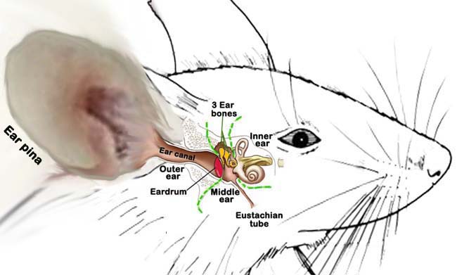
Figure
3.
Schematic
illustration
of
the
mouse
ear.
The
green
dash
lines
depict
the
anatomical
regions
of
the
ear.
The
eardrum
is
accentuated
in
red.
The
3
ossicles
or
ear
bones
(highlighted
in
orange)
include
malleus,
incus,
and
stapes.
(Not
drawn
to
scale.)
Testing
with
MT10
tympanometer
In
a
quite-noise
protected
procedure
room,
the
mobility
of
the
eardrum
is
tested
using
a
tympanometer
(see
Figure
1
above),
which
applies
a
small
amount
of
air
flow
producing
a
pressure
sensation
into
the
ear.
a.
Proper
ear
tip
selection
of
suitable
size
is
accomplished
before
testing
and
is
inserted
as
far
as
it
will
go
on
the
probe
tip
of
the
tympanometer.
b.
For
convenience
and
stable
positioning,
the
probe
tip
can
be
detached
from
the
main
housing
and
the
properly
sized
ear
tip
is
inserted
into
the
mouse
ear
canal
making
a
perfect
seal.
c.
To
facilitate
a
good
fitting
of
the
ear
tip,
the
ear
pinna
or
earlobe
is
pulled
out
and
the
ear
canal
is
straightened
out
during
insertion
of
the
ear
tip
into
the
ear
canal
opening
(see
Figure
4
below).
An
ear
tip
covered
with
petroleum
based
vaseline
may
be
necessary
to
obtain
a
perfect
seal,
assuring
that
the
opening
of
the
ear
tip
is
not
clogged
by
the
sealant
or
ear
wax
or
obstructed
by
the
wall
of
the
ear
canal.
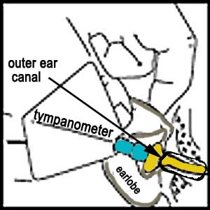
Figure
4.
Schematic
placement
of
the
tympanometer
ear
tip
within
the
ear
canal
opening.
d.
Once
the
ear
tip
is
properly
inserted
and
sealed
within
the
ear
canal
and
a
fixed
and
stable
position
is
applied,
the
test
is
automatically
started.
To
avoid
unwanted
movement
of
the
hand,
one
or
two
fingers
of
the
hand
holding
the
tympanometer
are
rested
on
a
fixed-stable
surface.
e.
Selected
ear
test
procedure
for
normal
tympanometry
is
conducted
first
in
one
ear
and
then
repeated
in
the
other
ear.
f.
After
each
test,
a
tympanogram
is
displayed,
graphically
charting
the
tympanic
membrane
compliance
under
changing
pressure
conditions
(see
Figure
2
above).
Tympanometric
parameters
(compliance,
volume,
gradient,
pressure)
are
measured
at
maximum
compliance
and
the
tympanograms
are
interpreted
according
to
manufacturer's
guidelines
and
other
clinical
validations
(i.e.
otoscopy,
ABR
threshold,
histology).
Data
collected
by
investigator
Tympanometric
parameters
for
both
right
and
left
ears:
compliance,
volume,
gradient,
and
pressure.