Gupta2 project protocol
Survey of vesico-ureteric reflux in newborns of 10 BXH recombinant inbred strains of mice (2011)
Gupta IRWith: Murawski I, Maina R, Myburgh D, Watt C, El Andalousi J
Project protocol — Contents
Workflow
Equipment, supplies, software
Reagents, solutions
Procedure: Dissection of newborn mice
Procedure: Screening newborn mice for VUR
Procedure: Kidney metrics (weight and planar surface area)
Definitions
Data
References
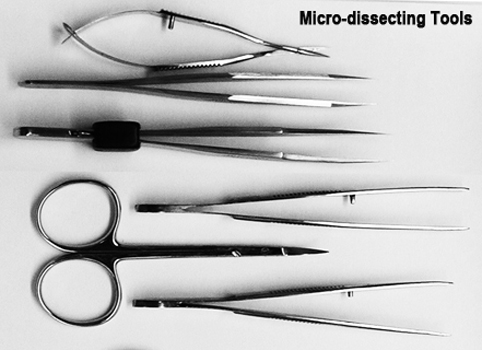
- Dissecting forceps (Dumont #5 Straight Tip Forceps-Rustless Dumoxel), Fig. 1
- Fine dissecting (iris) scissors (Sar-Med or Delicate Scissors 4.5" from VWR), Fig. 1
- Dissecting microscope (Leica MZ75) equipped with a camera (Leica Infinity 1)
- Software for moprhometric measurements (SPOT v.3.5.9)
- Timer
- Scale
- Metric ruler
- Eppendorfs (1.5 mL)
- Disposable petri dishes
- Surgical blade (Bard-Parker no.22 Stainless Steel Surgical Blade-single use)
- Beaker
- Needle (26 3/8 gauge intradermal bevel needle-Precision Glide)
- 60 mL syringe (Becton Dickinson)
- Ice
- 70% ethanol
- IV tubing that is at least 185 cm in length (Microbore Fat Emulsion Extension Set, 74 inch tubing, LifeShield, Hospira)
- Methylene blue (diluted to 1mg/mL in 1x sterile PBS)
Procedure: Dissection of newborn mice
a. Newborn mice are placed in a petri dish immersed in ice and allowed to become hypothermic for approximately 20 min.
b. Mice are weighed, sexed, and euthanized (decapitation).
c. With the aid of a dissecting microscope, a midline incision from the thorax to the abdominal cavity is made using fine iris scissors to expose the rib cage and underlying organs without disturbing the bladder.
d. Two additional incisions are made from the midline outwards or laterally to expose the kidneys and the urinary tract.
e. Using forceps and disposable surgical blade, excess tissues above the kidneys and surrounding organs are removed such that all that remains are the hindlimbs, spine, kidneys, and urinary tract.
f. For morphometric evaluation, bladder, ureters, and kidneys are dissected intact (in toto) from the body cavity by gently lifting the bladder and freeing the tissue from the rest of the body with fine dissecting scissors.Procedure: Screening newborn mice for vesico-ureteric reflux (VUR)
I. Pre-VUR assay essentials
a. Pre-set heights are marked on the wall adjacent to the dissecting microscope serving as a guide during the VUR assay. In marking the height of the column of dye a ruler is used to measure 5 cm intervals from the base of the microscope. Standardized heights/pressures intervals of 30, 60, 90, 120, and 150 cm relative to the microscope are used.
b. A 60 mL syringe is attached at one end of an IV tubing with a 26 3/8 gauge needle is attached to the opposite end.
c. The 60 mL syringe is then primed with methylene blue dye and allowed to flow, flushing the tubing and riding of any trapped air bubbles. Air bubbles are avoided by keeping the syringe filled with dye and by keeping it leveled with the base of the microscope. Raising and lowering the column is sufficient to start or stop the flow of dye.
d. The column of dye and needle are placed in a tall beaker for support until needed.II. Vesico-ureteric reflux (VUR) assay
a. The VUR assay is performed by two people using a dissecting microscope equipped with a digital camera.
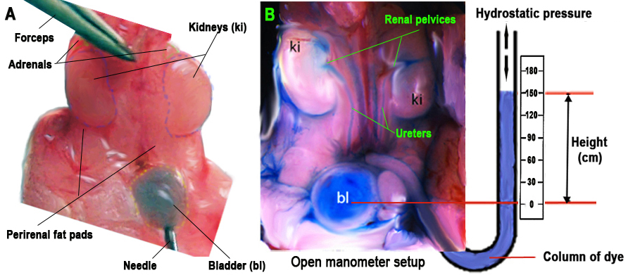
b. The first person is assigned the task of inserting one end of the tubing with a needle into the bladder while holding the bladder firmly with forceps under a dissecting scope, and making sure that the bladder is not punctured (Fig. 2A).
Figure 2. Schematic illustration of the open manometer setup. Panel A: Using forceps, the kidneys are held in place and a needle is inserted into the bladder as shown. Panel B: Once the needle is inserted, the column of dye is raised at specific intervals and the rate of dye flowing into the bladder is determined by the hydrostatic pressure exerted by the column of dye. When VUR is present, the dye can be seen flowing up the ureters into the renal pelvis. The hydrostatic pressure at which VUR is (or not) observed is measeured as the height of the column of methylene blue in relation to the level of the mouse. Pressure is recorded separately for the paired kidneys.c. The second person is given the task of manipulating the dye-filled column (syringe) by gradually moving upward from 30 cm to 150 cm at a rate of 30 cm every 5 s, for a total of 30 s. Upon reaching the maximum height (pressure) of 150 cm relative to the dissecting microscope, it is held in place for 10 s. At this height the hydrostatic pressure is at its maximum and the flow of dye into the bladder is at its greatest.
d. The first person then observes and records whether any dye flows retrogradely from the bladder into the ureters and/or kidneys.
e. At 150 cm the dye readily exits via the urethra. The height of the column concomitant with dye exiting the urethra is recorded as the voiding pressure. The voiding pressure varies between each mouse, and usually occurres between 30 and 60 cm (average 45 cm).
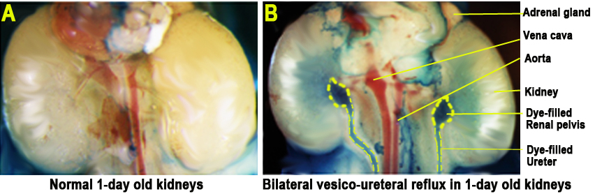
f. The height of the column concomitant with dye observed going up the left and/or right ureter and/or kidney is recorded as the VUR pressure. Since some mice exhibit unilateral VUR while others exhibited bilateral VUR, it is recorded separately for left and right ureter and kidney (Fig. 3A and 3B).
f. Following the VUR assay, the column of dye is lowered before the needle is retracted from the bladder to avoid excess dye on the kidneys, which are removed using fine forceps for further analysis (see below).
g. Scissors, forceps, and petri dish are cleaned with 70% ethanol in between each mouse tested for VUR.
h. When all mice have been tested, the IV tubing and the 60 mL syringe are rinsed with dH2O and reused. All needles and blades are disposed of in designated containers.
Figure 3: Newborn kidneys. Panel A: Normal ureters and kidneys. Panel B: Bilateral vesiculo-ureteral reflux in newborn mice. Renal pelvices and ureters oulined in yellow dashed lines.Procedure: Kidney metrics (weight and planar surface area)
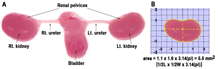
a. The kidneys are positioned lateral-medial as shown in Fig. 4A, then digitally photographed.
b. The paired kidneys are seperated and weighed.
c. Morphometric evaluation of kidney planar surface area is performed on digitized kidney images using SPOT (v.3.5.9) software after proper calibration (Fig. 4B).
d. Kidney planar surface area and weight measurements are normalized to body weight to account for any differences in gestational age in newborn mice.
e. Forceps and scissors are cleaned with 70% ethanol after each use.
Figure 4: Schematic layout of the kidneys for morphometric evaluation. Panel A: Bladder, ureters, and kidneys en bloc are placed on a petri dish, separated, and positioned in a lateral-medial view for digital photography. Panel B: Measuring kidney area using simple graphical axis (arbitrary scale for illustration).vesico-ureteric reflux (VUR): the retrograde flow of urine (or dye) from the bladder up the ureter, and potentially up to the renal pelvis into the kidney
vesico-ureteric reflux index: scored with up to a maximum 150 cm of dye hydrostatic pressure and according to the following degree of reflux involvement
0=no reflux
1=mild (only 1 ureter involved)
2=mild-moderate (both ureters)
3=moderate (1 kidney)
4=moderate-severe (1 kidney + 1 ureter)
5=severe (both kidneys)
Data collected by investigator
- body weight
- urethral hydrostatic pressure
- vesico-ureteric reflux (Supplementary data)
- kidney planar surface area (actual and adjusted for body weight)
- kidney weight (actual and adjusted for body weight)
MPD calculated
• vesico-ureteric reflux index (see scoring definitions above)