Handel1 project protocol
Male reproductive parameters in 14 inbred strains of mice (2010)
Handel MWith: O'Brien M
Project protocol — Contents
Workflow and sampling
Equipment and consumables
Procedures
Data
Definitions
References
Workflow
Mice are sacrificed by cervical dislocation Testes and seminal vesicles removed and weighed Epididymides are removed and processed for sperm count • Dissecting instruments: scissors, 2 forceps, scalpels
• Balance scale
• Inverted microscope
• Petri dishes (35 mm)
• Minimum essential medium (MEM, BSA 3 mg/mL)
• Ependorf tubes
• Pipette tips
• Automatic dispensers, pipettes
• Water sterile
• Hemocytometer
I. Organ weight measurement
a. For each male mouse 1 mL of media (MEM BSA 3 mg/mL) is pipetted into a small petri dish and warmed to 37°C.
b. 8-10 wk old male mice are sacrificed via cervical dislocation by fully trained personnel and subsequently weighed for body weight .
c. The entire male reproductive organs are exposed via an abdominal incision (see exteriorized organs below, Figure 1).
d. Both the left and the right testes are dissected and weighed together.
e. The seminal vesicles, with closely adjacent smaller coagulating glands, (see Figure 1 below) are also dissected and weighed.
f. The cauda (tail) of both (right and left) epididymides are carefully dissected, removed and placed in a petri dish prepared above (see step "a") for subsequent processing and sperm counting.
Figure 1. Schematic illustration of the male mouse reproductive organs.
II. Counting sperm
a. Both the left and the right cauda epididymides are incised, and the sperm is allowed to swim for 15 min out into the medium and form a suspension.
b. A 1:10 dilution is made by adding 90 µL of water to 10 µL of sperm suspension aliquoted into an ependorf tube.
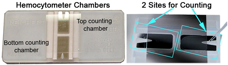
c. Sperm counts are obtained twice, each time using 5 squares taken from each of the 2 sites in the hemocytometer.
d. Average of the 2 counts is calculated and recorded.
Figure 2. Hemocytometer counting chambers.Data collected by investigator
Sperm count, seminal vesicle and testicular weights (both left and right)
Sperm count = Dilution x (Count in 5 squares) x 0.05 x 10⁶.
References
Lessard C, Pendola JK, Hartford SA, Schimenti JC, Handel MA, Eppig JJ. New mouse genetic models for human contraceptive development. Cytogenet Genome Res. 2004;105(2-4):222-7.
PubMed 15237210