Donahue8 project protocol
Plasma immunoglobulin concentrations in C57BL/6J-Chr#PWD/Ph/ForeJ mouse chromosome substitution strains (2011)
Donahue LWith: Morgan J
Project protocol — Contents
Workflow and sampling
Equipment and supplies
Reagents and solutions
Procedure for measuring plasma immunoglobulins
Definition
Data
ReferencesWorkflow
Mice are brought from the mouse room to the procedure room and acclimated before blood collection After acclimation, eyes are locally anesthetized for retro-orbital bleeding Whole blood collected into microcentrifuge tubes with EDTA Blood samples processed for plasma sample collection and frozen until needed Frozen plasma samples packed in dry ice and sent away for IgA, IgG, and IgM concentration determination
- blood collection kit (microcentrifuge tubes, heparinized hematocrit tubes)
- centrifuge
- cryostat tubes
- -80°C freezer
- 96-well ELISA plates
- spectrophotometer
- topical anesthetic Tetracaine
- EDTA anticoagulant
- mouse IgA, IgG, and IgM ELISA Kit (GenWay Biotech, Inc., San Diego, CA)
- plasma diluent (0.9% saline)
- dry ice
Acclimation to test conditions
Blood collection is conducted during the light phase between 08:00 and 12:00 h. Mice are transported from the mouse room into the procedure room and are allowed to acclimate for at least 1 h.
Procedure for measuring plasma immunoglobulins
a. After administering Tetracaine on the eye as a topical anesthetic (volume ~200 µL), peripheral blood is obtained by retro-orbital bleed.
b. Approximately 250 µL of whole blood is collected in tubes with EDTA anticoagulant.
c. Blood samples are centrifuged for 3 min, and plasma is pipetted into cryostat tubes and frozen at -80°C until shipped from JAX to GenWay on dry ice.
d. Measurement of mouse plasma IgA, IgG, and IgM is performed by GenWay.
e. Each sample is diluted first before conducting ELISA on a 96-well plates.
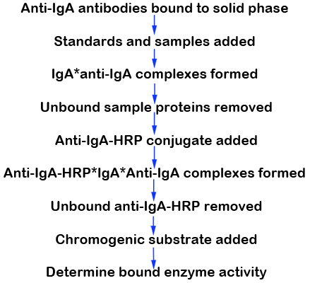
f. The standard curve is reported in ng/mL and multiplied by the dilution factor to convert to µg/mL.GenWay protocol for immunoperoxidase assay for determination of mouse IgA, IgG, and IgM from plasma (Fig. 1)
1. The principle of the double antibody sandwich ELISA is represented in Figure 1.
2. In this assay the immunoglobulins present in plasma samples reacts with the anti-IgA, IgG, or IgM antibodies which have been adsorbed to the surface of polystyrene microtitre wells.
3. After the removal of unbound proteins by washing, anti-IgA, IgG, or IgM antibodies conjugated with horseradish peroxidase (HRP) are added. These enzyme-labeled antibodies form complexes with the previously bound IgA, IgG, or IgM.
4. Following another washing step, the enzyme bound to the immunosorbent is assayed by the addition of a chromogenic substrate, 3,3’,5,5’-tetramethylbenzidine (TMB).
5. Since the quantity of bound enzyme varies directly with the concentration of IgA, IgG, or IgM in the samples, the concentration of IgA, IgG, and IgM is reflected at 450 nm absorbance.
6. The quantity of IgA, IgG, and IgM in the plasma sample is interpolated from the standard curve constructed from the standards, and then corrected for sample dilution.
Figure 1. Immunoperoxidase assay for determination of IgA mouse plasma.ELISA: enzyme-linked immunosorbent assay that uses anti-immunoglobulin antibodies bound to solid-phase enzyme immunoassay (EIA) to detect the presence of immunoglobulins in plasma sample.
The standard curve is reported in ng/mL and multiplied by the dilution factor to convert to µg/mL.
Data collected by investigator
- plasma immunoglobulin concentration
- IgA
- IgG
- IgM